Organism Microscope: What It Is, What It Shows and How to Choose one
A drop of pond water looks empty. Under the right microscope, it turns into a crowded world of swimming, feeding and dividing life.
That is the promise of an organism microscope. Whether you are setting up a school lab, starting a home hobby or equipping a microbiology bench, the real question is the same: which instrument will actually show you living organisms clearly?
This guide answers that from the ground up.
What Is an Organism Microscope?
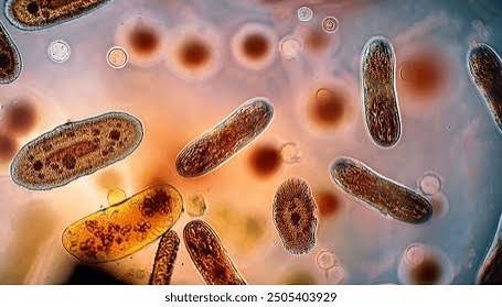
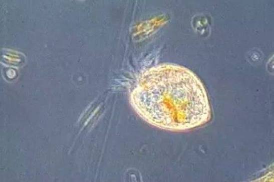
An organism microscope is a microscope used to observe living or preserved microorganisms and cells, such as protozoa, algae, yeast, fungi, plant cells and stained bacteria. In practice, it is usually a compound light microscope with magnification from about 40x to 1000x.
The phrase describes a purpose, not a separate product category. In catalogs you will find the same instruments listed as biological microscopes, microbiology microscopes, bright-field microscopes or student microscopes.
So when a listing says “organism microscope”, the label tells you little. The optics, light and build tell you everything.
Why Observing Organisms Matters
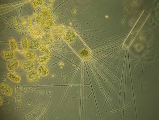
The scientific study of microorganisms began when Antonie van Leeuwenhoek looked at them through simple single-lens microscopes in the 1670s. Robert Hooke’s earlier work on cells and Louis Pasteur’s later work on microbes built on that same habit of looking closely.
Today the same skill supports many fields:
- Education: Students see cells and protists instead of memorizing diagrams.
- Clinical and diagnostic work: Smears and stains, including the Gram stain, begin with a microscope.
- Water and environmental checks: Algae and protozoa hint at what is living in a sample.
- Fermentation and food work: Yeast and culture health can be checked at a glance.
- Research: Cell and microbe studies still start with observation.
Who Needs One, and When
Different users need very different instruments.
- Children and hobbyists: An entry-level model for pond water, onion skin and yeast.
- Schools and colleges: Durable binocular compound models that survive daily student use.
- Labs and clinics: Higher-grade optics, often with a camera port.
- Researchers working with live cells: Phase contrast or inverted systems, which show transparent living cells without heavy staining.
If you want to watch live, unstained cells in detail, plan for phase contrast. If you mainly want to find protozoa and algae in pond water, a good standard compound microscope is enough.
How an Organism Microscope Works
A compound microscope bends light through two lens systems in sequence. Understanding the path makes troubleshooting much easier.
- Light source: An LED, or a mirror in simple models, lights the specimen from below.
- Condenser and iris diaphragm: These shape the light and control contrast.
- Objective lens: The first magnification, usually 4x, 10x, 40x and 100x.
- Eyepiece (ocular): The second magnification, usually 10x.
- Focus knobs: Coarse focus finds the image, and fine focus sharpens it.
Total magnification is the objective power multiplied by the eyepiece power. A 40x objective with a 10x eyepiece gives 400x.
Most good microscopes are parfocal, which means the image stays roughly in focus as you switch objectives. That makes moving up in power fast and safe.
The image is also inverted. Move the slide left and the image appears to move right. It surprises every beginner once.
Magnification Is Not the Same as Resolution
Resolution is the ability to separate two close points. It depends on the objective’s numerical aperture (NA) and the quality of the glass, not on the number printed on the box.
This is why a cheap model labeled 1500x or 2000x can still give a blurry image. Past the useful limit of the optics, you only get a bigger blur. That is empty magnification.
When comparing models, look at objective quality, NA values if listed, and whether the lenses are glass. These matter more than the headline figure.
What You Can See at Each Magnification
The table below is a practical guide. Sizes vary by species, so treat it as a starting map.
| Magnification | What typically becomes visible |
| 40x | Algae colonies, larger protozoa and tiny pond animals in context |
| 100x | Paramecium, Euglena, Volvox and yeast clusters begin to show clearly |
| 400x | Cell structures, moving protozoa, rotifers, plant cells and fungal strands |
| 1000x (oil immersion) | The shape of bacteria, mostly after staining |
Bacteria are the common trap. At 400x you may notice that something is there, but you will not reliably see its shape. For that you need a 100x oil immersion objective, good light and usually a stain such as methylene blue or a Gram stain.
First organisms worth finding
Start with subjects that are easy to find and forgiving to focus.
- Pond water or hay infusion: Paramecium and other ciliates.
- Green pond water: Euglena and Volvox.
- Aquarium plant (Elodea): Cell walls and moving chloroplasts.
- Yeast in warm sugar water: Budding cells.
- Onion skin: A classic, clean first slide.
- Cheek cells with methylene blue: A good stained-slide exercise.
Types of Microscopes for Observing Organisms
The right type depends on what you want to watch and how you want to record it.
| Type | Best for | Strength | Limit |
| Monocular compound | Beginners, kids | Low cost | Eye strain in long sessions |
| Binocular compound | Classrooms, serious hobbyists | Comfortable viewing | Higher price |
| Trinocular compound | Labs, documentation | Camera port | Costs more |
| Digital microscope | Sharing on a screen | Easy recording | Often weaker optics at the same price |
| Stereo (dissecting) | Insects, leaves, larger organisms | 3D view, no slide needed | Low magnification |
| Phase contrast | Live, transparent cells | Contrast without stains | Specialized and pricier |
| Inverted | Cells in dishes | Objectives sit below the sample | Mostly research use |
Compound vs. stereo for organisms
A compound microscope views thin, transparent samples at high power. A stereo microscope gives a 3D view of larger, solid objects at low power. For microbes, choose compound. For beetles, seeds or moss, choose stereo.
Monocular vs. binocular
Monocular is fine for short, casual sessions. Binocular is easier on the eyes and worth the extra money if the microscope will be used daily or by several students.
Optical vs. digital
A digital microscope is convenient for projecting images to a class. At similar prices, an optical compound microscope generally resolves more detail. Many people pair an optical model with a camera adapter or a trinocular port.
Features That Actually Matter
When you compare models, put these in order.
- Glass objectives: Achromatic is the common standard. Plan objectives give a flatter field and cost more.
- Objective range: 4x, 10x, 40x and, if you need bacteria, a 100x oil objective.
- LED illumination: Cool, long-lasting and consistent. A mirror works off the grid but is harder to control.
- Condenser with iris diaphragm: This is essential for contrast. Many beginners overlook it.
- Mechanical stage: It makes moving a live sample far easier.
- Metal frame: A stable frame keeps the image from shaking at high power.
- Spare parts and support: Replacement LEDs, objectives and a warranty matter over years.
- Field of view and working distance: Useful for comparing models once the basics are right.
For consumer sales, look for recognized safety marks such as CE and RoHS compliance. Established makers include Olympus (now Evident), Nikon, Zeiss, Leica, AmScope and Motic. Check current model lines before you buy, since catalogs change often.
How to Choose: A Simple Decision Path
Work through these steps in order.
- Name the organisms. Pond water and yeast need about 400x. Bacteria need 1000x with oil.
- Decide live or stained. Live and unstained work is best with phase contrast. Stained work suits standard bright-field.
- Pick the viewing style. Short sessions suit monocular. Long sessions suit binocular.
- Decide on recording. If you need photos or video, choose a trinocular model or a camera adapter.
- Check the build. Metal frame, glass lenses, smooth focus and a real diaphragm.
- Set a budget and compare two or three models in that range.
Quick match by user
- Child or casual hobbyist: Entry-level monocular, 40x to 400x, LED.
- Student or school lab: Binocular compound, 40x to 1000x, mechanical stage.
- Home microbiology enthusiast: Binocular with a 100x oil objective and a camera adapter.
- Clinic or lab: Trinocular, plan objectives, strong illumination.
- Live-cell researcher: Phase contrast or inverted system.
What It Costs
Prices vary by country, import duty, exchange rate and retailer, so treat these as rough bands and confirm current prices before buying (Source: manufacturer and retailer listings, 2026).
| Category | Rough range (USD) |
| Starter and kids’ models | About 30 to 100 |
| Student binocular | About 150 to 400 |
| Lab-grade compound | About 500 to 2,000 |
| Phase contrast or inverted | About 1,500 and above |
Used or refurbished instruments from established brands can be good value. Inspect the lenses for fungus, scratches and haze, and make sure the focus moves smoothly.
Also factor in service. Spare LEDs, shipping costs and local repair availability can change the real cost of ownership.
How to View Pond Water: Step by Step
- Collect a sample. Take water from near plants or the pond edge, or soak hay in water for a few days.
- Make a wet mount. Place one drop on a clean slide.
- Add a cover slip. Lower it at an angle to avoid air bubbles.
- Start at the lowest objective. Use 4x or 10x and focus with the coarse knob.
- Adjust the light. Close the iris diaphragm slightly until contrast improves.
- Step up in power. Switch to 40x and use only the fine focus.
- Slow fast swimmers. A drop of methyl cellulose, or a few cotton fibers, holds them in view.
- Record and clean up. Photograph through the eyepiece or camera port, then wipe the objectives with lens paper.
Why You Can’t See Anything: Troubleshooting
Most problems are simple.
- Blank bright field: The sample is probably not under the lens. Return to 4x and look around the slide.
- Washed-out image: Close the diaphragm a little. Wide-open light kills contrast.
- Blurry at 40x: The slide may be upside down, or the cover slip too thick.
- Nothing moves: The drop may be too thin, or the sample too clean. Try hay infusion.
- Fast blur of movement: Use methyl cellulose or a lower power.
- Dark image: Check the LED, raise the condenser and open the diaphragm slightly.
- Dirty haze: Clean objectives with lens paper, and never with a cloth or tissue.
Live, Stained and Fixed Specimens
Live specimens show movement and behavior, but they are hard to keep still and low in contrast. Stains such as methylene blue add contrast and are common for cells and bacteria, but stained samples are usually no longer alive. Fixed or prepared slides last for years and are ideal for teaching.
For live, unstained cells, phase contrast turns small differences in thickness into visible contrast. This is why it is valued in cell biology.
Electron microscopes can show far more detail, but preparing samples for them kills the specimen. Live work stays with light microscopy.
Common Mistakes
- Buying on magnification alone. Check optics and build first.
- Ignoring the diaphragm. Poor contrast is often a lighting problem.
- Forcing the focus. At high power, wrong focusing can crack a slide or scratch a lens.
- Putting oil on the wrong objective. Immersion oil belongs only on the 100x oil objective.
- Leaving oil behind. Clean it after each use to protect the coatings.
- Storing it uncovered. Dust and humidity invite lens fungus.
Safety and Care
Never use direct sunlight as a light source. It can cause serious eye damage.
Stick to safe, familiar samples at home: pond water, hay infusion, yeast, plant cells and cheek cells. Do not culture unknown bacteria in a household setting. Wash your hands after handling samples, and keep samples away from food areas.
For care, use lens paper, keep the dust cover on, lower the stage after use and add silica gel in humid climates.
Alternatives
- Stereo microscope: Better for insects, leaves and minerals.
- Digital microscope: Good for classroom screens.
- Pocket or handheld microscope: Handy for fieldwork, with lower resolution.
- Prepared slide sets: A good way to learn without handling live samples.
- Lab or school access: Useful if you only need occasional viewing.
Frequently Asked Questions
1.What is an organism microscope?
It is a microscope, usually a compound light microscope, used to observe microorganisms and cells such as protozoa, algae, yeast and stained bacteria.
2.Which microscope is used to see living organisms?
A compound light microscope shows most live microorganisms in a wet mount. Phase contrast microscopes show transparent living cells more clearly, and inverted microscopes suit cells in culture dishes.
3.Can you see bacteria with a school microscope?
Only with 1000x oil immersion and, ideally, a stain. At 400x you may notice bacteria are present, but not their shape.
4.What magnification do you need to see microorganisms?
About 100x to 400x for protozoa and algae. For bacteria, plan on 1000x with oil immersion.
5.Compound vs. stereo microscope for organisms: which is better?
Choose compound for microscopic organisms on slides. Choose stereo for larger, solid specimens such as insects and plant parts.
6.Why can’t I see anything moving on my slide?
The drop may be too clean, too thin or out of the field of view. Try pond water or hay infusion, start at low power and close the diaphragm slightly for contrast.
7.Do I need oil immersion?
Only for bacteria and fine cell detail at 1000x. Most pond-water viewing is done at 100x to 400x without oil.
8.Is a digital microscope good enough for organisms?
It is fine for sharing and recording larger or brighter subjects. For fine detail in microbes, an optical compound microscope usually performs better at the same price.
9.Is it safe to look at pond water at home?
Yes, with normal hygiene. Wash your hands afterward, avoid culturing unknown microbes and keep samples away from food.
Practical User Experience
This section describes what people typically notice when they first use an entry-level or student compound microscope. It reflects how these instruments generally behave, not one specific model.
The first surprise is how empty the view looks. Finding the sample at 4x or 10x takes patience, and the image often stays flat until the diaphragm is closed a little. Once contrast improves, the picture changes quickly.
Live samples are the second lesson. Pond-water organisms swim out of view in seconds, so a drop of methyl cellulose or a few cotton fibers makes a real difference. Hay infusion is usually richer than plain pond water.
Handling matters more than expected. A mechanical stage makes moving the slide far easier than doing it by hand, and a metal frame keeps the image steady at 400x. Lighter plastic bodies tend to wobble when the focus knob is touched.
Bacteria are where expectations meet limits. At 400x the field shows that something is there, but its shape needs oil immersion and a stain, and those steps take time to learn.
Overall impression: a well-built binocular compound microscope handles protozoa, algae and yeast reliably and rewards slow, careful setup. Budget models can still teach the basics, but they limit detail and comfort, so treat them as a starting point.
Conclusion
An organism microscope is best understood as a compound light microscope matched to the organisms you want to see. For protozoa, algae and yeast, a well-built 400x model is plenty. For bacteria, you need quality 1000x optics, oil immersion and a stain.
Buy for resolution, light control and build quality, not for the biggest number on the box. Start with pond water and yeast, learn to use the diaphragm, and move up in power one step at a time.
Unfulfilled Points: An expert reviewer line and a “how we tested” note were not added because they require real credentials and hands-on input. Exact ISO standard numbers and brand model details were left general because they need verification, and structured data is a publishing step rather than article text.
These information are gathered from different surveys, medical tests and tech giants like Techradar and more. Our top priority is to provide you valuable information about Organism Microscope .
For More Details About Different Digital Microscopes Click Here