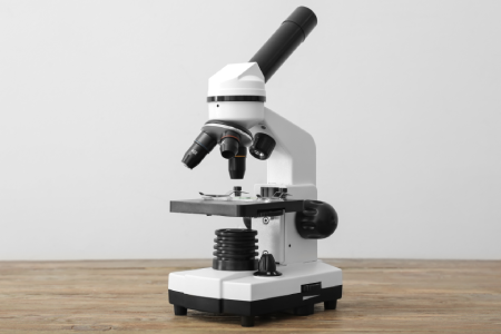
Compound microscopes are instruments that employ numerous lenses to enhance the dimensions of tiny objects, generally from 40x up to 1000x, meaning that even though structures appear invisible to the naked eye, bacteria, such as cells that are thin in sections, can be seen and observed.
Cells in thin tissue sections are visible and observable. This is the typical microscope that can be found in labs for clinical use, classrooms, as well as research settings all over the world.
If you’ve ever viewed an instrument in biology classes or even had blood drawn to test your lab, you likely used a compound microscope at some point in the process. It’s among the most frequently utilized scientific instruments; however, the majority of people buying a microscope at first don’t know what the difference is between a toy version and a more expensive lab-grade version, or whether they actually require a compound microscope in comparison to something else.
This guide will explain the basics of what a compound microscope is, how it operates with the key components, as well as the different types of models available and a simple approach to choosing which is best for your particular needs, regardless of whether it’s an at-home science kit or a lab at a university or a smaller lab.
What Is a Compound Microscope Exactly?
The expression “compound” refers to the lens, not the size of the device. A compound microscope consists of at least two lenses, working together, comprising an optical lens situated near the specimen and an optical lens (the eyepiece) located near your eyes. Light travels over the subject; the objective lens magnifies it, then it is magnified again by the eyepiece.
This magnification in two stages is what can make a compound microscope distinct from a standard magnifying glass, which is based only on one lens and is capped at a low magnification. It’s also different than stereo microscopes, which make use of two different optical pathways to provide the illusion of 3D views of an object, rather than a flat and highly magnified image of an incredibly thin object. Compound microscopes fall into the class of microscopes that also includes optical microscopes as well as optical microscopes since they are based on visible light, which passes through (or bounces off) the subject, instead of electron beams or any other imaging methods.
How a Compound Microscope Works
The mechanisms are much more straightforward than they appear. Light — whether it’s natural or derived from an LED or halogen bulb integrated inside the base flows through the object at the bottom of the platform, into the lens of the objective, and upwards through the body tube until it reaches the eyepiece.
Magnification can be explained using two figures.
Total Magnification = Objective Lens Power x Eyepiece Lens Power
A 40x objective combined with an eyepiece magnifier of 10x is equivalent to 400x, but most compound microscopes allow users to adjust magnification without changing instruments – typically 4x 10, 10x 40x, or 100x objective lenses are interchangeable on turrets for increased magnification without switching instruments.
One thing that is a source of confusion is that A compound microscope image can be reversed and inverted compared to the real orientation of the object. This is because light bends when passing through the convex lens. This is an optical phenomenon that is normal and not a flaw within the microscope.
The Parts That Actually Matter
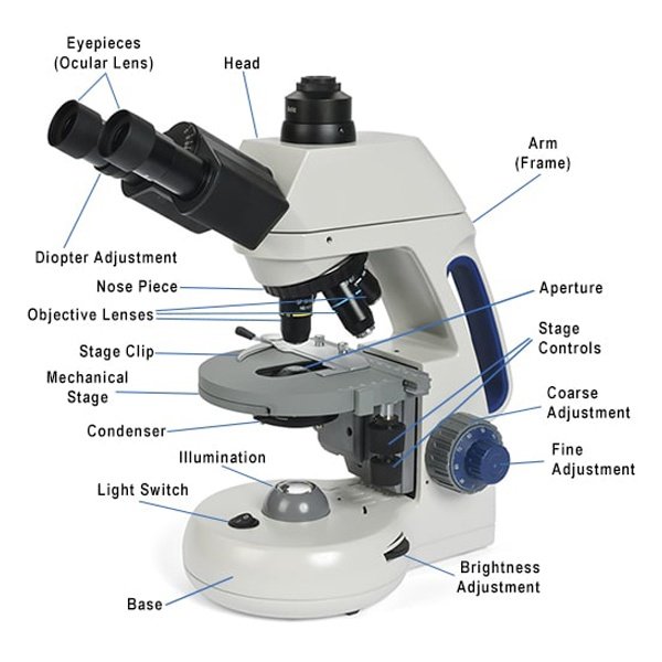
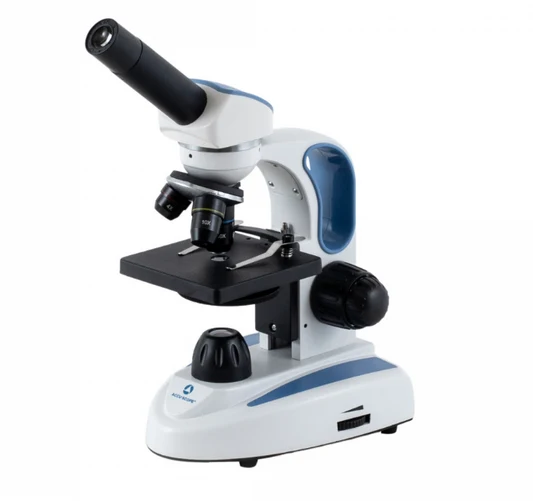
Each compound microscope, no matter the cost, is built on the same basic elements:
Part What It Does
| Eyepiece (ocular lens) | It is the second step of magnification. You can see what you are looking through |
| Objective lenses | Offer the initial stage of magnification, mounted on the nosepiece and rotating. |
| Nosepiece | A rotating turret which holds and changes between the lenses of the objective |
| Stage | Stage Flat platform which holds the slide in its entirety, typically with clips to help keep it stable. |
| Condenser | The lens focuses light on the object before it is able to reach the lens. |
| Diaphragm | An adjustable opening controls how much light enters |
| Fine and coarse focus knobs | Coarse to get a rough focus; fine for sharpening the final image |
| Arm and base | The structural frame that holds everything together. |
An important design aspect to be aware of before purchasing the best microscopes: they have been designed to be parfocal, which means that the object remains in focus when you switch between the objective lenses. Less expensive models usually aren’t, and you’re forced to focus on the spot every time you alter the magnification- an insignificant issue that can become a major annoyance after frequent usage.
Types of Compound Microscopes
There are many different compound microscopes that are constructed the same way, and the differences in their construction are more than buying guides acknowledge.
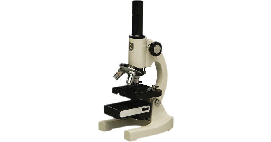
| Monocular Eyepiece: | One the simplest and least expensive, most commonly used in schools and at entry-level. |
| Binocular | two eyepieces. Better suited for longer viewing sessions and is standard for most labs |
| Trinocular | binocular view and a third port to connect a camera It’s perfect for recording as well as teaching and sharing pictures. |
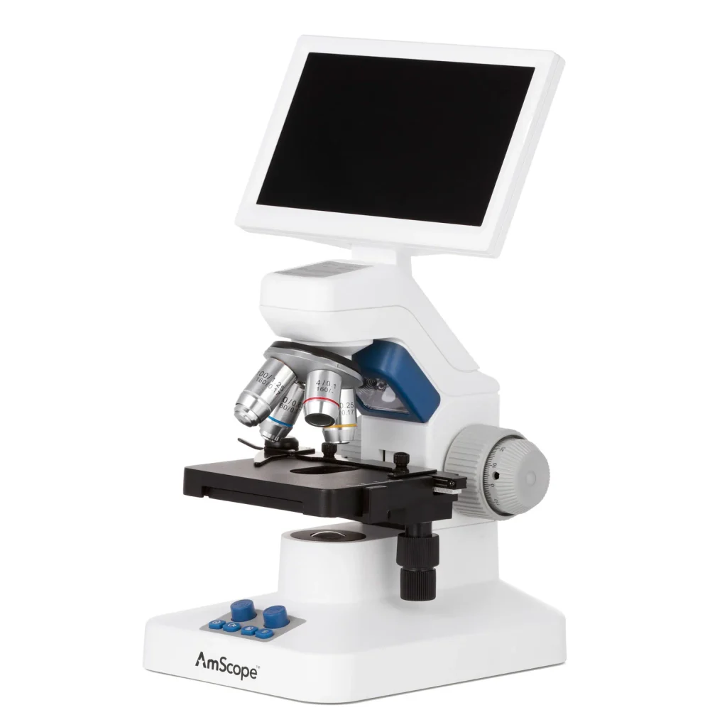
| Digital | Eyepiece and Screen This is a method that uses an internal camera as well as a screen, instead of an eyepiece. It often used in classes to allow all students to be able to view the same image at once. |
| Phase-contrast | enhances contrast in samples which resemble living cells but without staining. This could increase contrast while putting the specimens at risk. |
| Fluorescence | utilizes fluorescent light and markers to show specific structures. This is a common practice in more sophisticated research. |
For most hobbyists and students, the binocular compound microscope will cover the majority of usage scenarios. Fluorescence and phase-contrast models are tools with specialized features that many users will never require.
Compound is different from. Stereo Vs. Simple Microscope
This is among the most frequent sources of confusion. It’s important to get it cleared up before you purchase the incorrect instrument.
Feature Compound Microscope Stereo Microscope Simple Microscope
| Lens system | Multiple lenses, two-stage | Two distinct optical paths | Single lens |
| Magnification range | 40x-1000x | 10x-40x | Up to ~20x |
| Image | Flat (2D) which provides | Highly precise 3D | 2D, low-detail image |
| Best for | Blood, cells, bacteria and tissue | Circuit boards, whole insects and rocks | magnification of objects |
If you’re looking to observe the individual cells or microorganisms in your environment, then you’ll need the compound microscope. If you want to study entire objects such as a coin, an insect, or even a circuit board in three dimensions, the stereo microscope can be described as the best device, and it will fail you since it isn’t able to be focused on anything that has the depth of a real object.
What a Compound Microscope Can — and Can’t — Do
The majority of buying guides do not mention this point; however, it is important. The optical microscope is limited to the frequency of visible light, which puts a high limit on the amount of detail they are able to capture -regardless of how high the magnification is.
In the real world, A compound microscope will clearly demonstrate bacteria, usually at 1000x magnification with an oil-immersion lens. What it is unable to show is viruses, as they are below the resolution limits for light microscopy. The ability to view viruses requires an electron microscope. It is a fundamentally different and much more expensive technique.
It is an extremely important thing to consider prior to buying a product when you want to achieve the level of detail required by a virus; no compound microscope, regardless of how costly, will help you get there.
Who Uses Compound Microscopes, and For What
- Teachers and students in biology classrooms, homeschooling programs, and university classes typically study animal and plant cells, onion skins, as well as pond specimens.
- Medical and clinical personnel -Blood smear analysis, urinalysis, pathology, and smear analysis work in hospitals as well as diagnostic laboratories.
- Microbiologists -Identification of microorganisms and bacteria and research.
- Veterinarians (fecal tests and scrapings of the skin.
- Hobbyists studying the pond’s water, plant cells, or household samples due to curiosity.
The device can be used for anything from a basic $60 classroom kit up to an expensive research setup of $10,000 or more, and the best option is largely dependent on the category you are in.
How to Select the Appropriate One ?
Before considering features, start by considering your use case, as this helps narrow the options quickly.
If you’re purchasing for students or in a homeschool environment: A monocular or basic binocular with 40x-400x magnification will almost always suffice. There is no need for phase-contrast or oil immersion to see onion cells or pond water.
If you’re purchasing for a diagnostic or clinical laboratory: Go binocular at least, and seriously contemplate trinoculars if documentation or teaching is a part of the process. You’ll need a 100x oil immersion objective for blood work and bacterial analysis, as well as reliable, consistent lighting with LEDs.
If you’re purchasing to conduct research: Trinoculars that integrate cameras are almost essential, and a phase-contrast or fluorescent capability could be needed based on the specific field of study you’re working in.
Certain practical aspects are applicable to all tiers:
- Glass optics are superior to plastic lenses in terms of sharpness and endurance, particularly for things that are used frequently.
- Metal frames made of all-metal stand up much better than plastic-bodied models when used in classroom or shared lab settings.
- Illumination from LED is now the norm. It’s brighter, more stable, and less expensive to maintain than older halogen bulbs.
- Design with parfocal can save you time and stress in the event that you’re frequently switching magnifications.
What Compound Microscopes Typically Cost ?
Prices vary significantly based upon the specs and quality. These are averages from the market, so you must verify the prices at the retailers you are considering before buying, because prices fluctuate over time..
Tier Typical Range Fits Best
| Tier | Typical price Range | Fits Best |
| Entry-level/student | $50-$150 | Homeschool, middle/high school |
| Mid-range binocular | $150-$500 | Labs at colleges as well as serious hobbyists |
| Professional binocular/trinocular | $500-$2,000+ | Small research labs, clinical laboratory environments |
| Advanced (phase contrast/fluorescence | $2,000-$10,000+ | Universities, research specialized |
The most common mistake is purchasing above your current level of education. A homeschool parent shouldn’t be looking for a trinocular research system that costs $2,000, or a clinical lab shouldn’t be relying on a toy model of $60 for diagnostic work.
Using a Compound Microscope the Right Way
- Set your microscope on the surface of a solid object, after that, turn to the light source.
- Make sure to clean the lens and slide by using lens paper prior to use.
- Set it at the middle of the stage and ensure it is secured using stage clips.
- Begin by using the lowest-power lens (usually 4x)
- Make use of the knob for coarse focusing to get the object into sharp focus.
- Turn the fine-focus knob to increase the sharpness of the image.
- . Adjust the diaphragm so it can manage light levels and enhance contrast, then gradually increase magnification by using the fine adjustment knob at every stage.
- To do this step-wise: progressively focus and zoom using the fine knob. -.
- For viewing 100x oil immersion, apply the oil directly onto the slide prior to turning the object into its desired position.
Common Mistakes to Avoid (Common Pitfalls of Software Testing)
- Beginning with High Power. Always begin on the lowest level; not jumping straight up to 100x or 40x makes it difficult to find your specimen and may cause cracks in the slide.
- The focus knobs are forced to move. If a knob refuses to move, stop and think about whether it’s not aligned correctly, and bending it could cause damage to the mechanism.
- The omission of cleaning the lens. A smudged lens is among the most frequently cited causes of blurred images and is the first thing you should check prior to concluding that the microscope is defective.
- Use the wrong cleaning substances. Never use household glass cleaner on your optics because it may damage the coatings of lenses. Make sure to use only the best lens paper and a lens cleaning solution.
- Storing it in a careless manner: Store a microscope using its lowest power objective and cover it with an enclosure to keep out dust.iterfuhrendeiterfuhrendeiterfuhrende
Frequently Asked Questions [ FAQs ]
1. Where can an optical microscope compound be employed?
These devices are able to see tiny specimens which would otherwise be difficult for our eyes to discern, like blood, cells, bacteria samples, and thin tissue slices in education, medicine and research settings.
2. What is the highest magnification that can be obtained through an optical compound microscope?
Compound microscopes which are common in their design usually have the capability of 1000x total and 100x lens magnifying oil as well as 10x magnification for the eyepiece, and so on.
3. What are the most important ingredients of an effective compound microscope?
The eyepiece, nosepiece goal lenses diaphragm and condenser. Light source, as well as coarse and fine focus knobs comprise the main components.
4.How does a compound microscope create inverted images? This phenomenon occurs due to how light bends when passing through its curved lenses; it should not be considered an issue with your microscope!
5.Does an image from a compound microscope exist in three- or two-dimension? No. Because its view lies across a thin specimen rather than on its surface, two dimensions are all that’s available here.
6.Do compound microscopes detect viruses? No. The size of viruses is less than the resolution of visible light; therefore, they’re not something an optical microscope can detect, which is why you need the use of an electron microscope.
7.What is the price of an expensive basic microscope?This makes them considerably more comfortable during long viewing sessions.
Conclusion
A compound microscope is able to earn its position in the list of most beneficial tools in the fields of science and education since it can do one thing very effectively: it makes an invisible image, ranging from one onion cell to an entire smear of bacteria. The key to choosing the best one isn’t to go after the most powerful magnification rating. It’s about making sure you match the model, type, quality, and price to the actual thing you’re going to examine. Make sure you’ve got it right, and even an entry-level model can last for a long time.
These information are gathered from different surveys, medical tests and tech giants like Techradar and more. Our top priority is to provide you valuable information about Compound Microscope .
For More Details About Different Digital Microscopes Click Here